
3D細胞培養專用HydroGel的專家,其主力產品VitroGel®完全無動物來源(Xeno-Free),最適合幹細胞或任何cytokine-sensitive細胞培養研究!
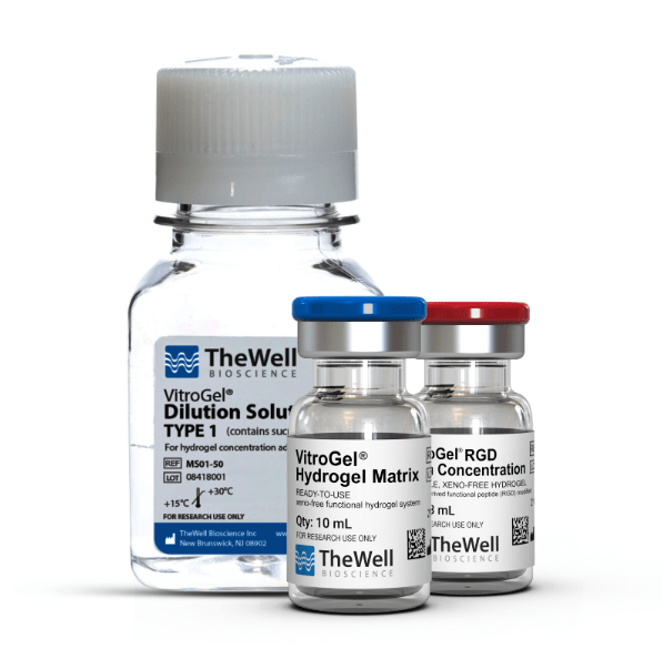
OSA 1777細胞株於VitroGel® 3D培養環境形成Spheroid
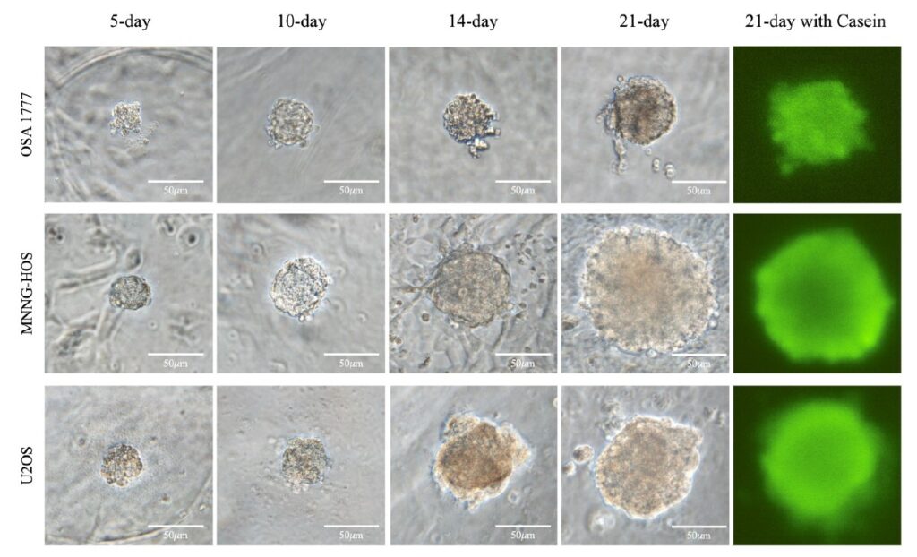
TheWell Cyto3D® Live-Dead Assay Kit
Versatile, live/dead cell viability analysis for 3D and 2D cell culture.
Cyto3D® 活/死 細胞檢測試劑盒
產品特色
- 適用於 3D 和 2D 細胞培養,包括類器官(organoids)、球體(spheroids)、幹細胞;兼容動物 ECM 和各種水凝膠系統。
- 採用雙重螢光顯像分析模式,能同時檢測活細胞與死亡細胞。
- 使用簡單、快速:無需預混合,可直接使用,一步染色完成。
- 染色均勻,影像清晰無背景干擾,適合高對比度成像。
染劑組成與機制
- 試劑盒含 吖啶橙 (Acridine Orange, AO) 與 碘化丙啶 (Propidium Iodide, PI),皆為核染劑,能結合核酸。
- AO 可進入活細胞與死細胞,將所有有核細胞染成 綠色。
- PI 僅能穿透膜受損的死亡細胞,使其呈現 紅色。
- 二者併用時,PI 會抑制 AO 的螢光,因此結果為:活細胞顯示綠色,死亡細胞顯示紅色。
- 不含細胞核的物質(如紅血球、血小板或細胞碎片)不會被檢測。
用途推薦
- 適用於細胞株、原代細胞及幹細胞的活性分析,尤其適合 3D 與 2D 培養系統。
- 可搭配螢光顯微鏡、流式細胞儀、微孔板讀取器或螢光細胞計數儀使用。
操作流程
- 將試劑恢復至室溫後使用。
- 加入 2 μL 試劑 至每 100 μL 的樣品中(可依實際水凝膠與培養液比例調整)。
- 在 37°C 下孵育 5–10 分鐘,即可進行細胞活性檢測。
規格與保存
- 試劑為預混 AO + PI 核染劑。
- 保存條件:2–8°C,避光儲存;保存期限約 15–24 個月。
- 出貨時可常溫運輸。
- 容量:1 mL,約可進行 500 次檢測(依 2 μL/100 μL 使用量計算)。
- 螢光參數:
- AO 激發/發射波長:約 494/517 nm,相容 GFP 濾片。
- PI 激發/發射波長:約 535/617 nm,相容 Texas Red 濾片。
應用案例
- 可用於 3D 水凝膠中腦膠質瘤細胞的活/死染色,並進行 z-stack 成像與 4D 重建分析。
- 在幹細胞球體中能進行穿透性良好的染色,清楚區分活細胞與死亡細胞。
Specifications
| Formulation | Premixed acridine orange (AO) and propidium iodide (PI), nuclear staining dyes |
| Use | Live dead cell viability analysis for 3D and 2D cell culture |
| Detection Method | Fluorescent |
| Excitation/Emission: | AO (494/517nm), PI (535/617nm) |
| Standard filters | AO (GFP), PI (Texas Red) |
| For use with (Equipment): | Fluorescence microscope, flow cytometer, microplate reader, fluorescence cell counter. |
| Storage | 2 to 8°C (Protect from light) |
| Shipping Conditions: | Ships at ambient temperature |
| Sizes | 1 mL |
| Number of reactions | 500 (at 2 µL per 100 µL) |
Data and References

Figure 1: Live-dead cell viability images: Intestinal organoids stained with Cyto3D® Live-Dead Assay Kit.
Intestinal organoids were cultured in regulated conditions for 5 days. Six microliters of Cyto3D® reagent were mixed with organoid culture media (each well includes 150 µL of organoid culture media and 150 µL of hydrogel volume). The mixture was incubated at 37°C for 10-15 min, and the cells were observed under a fluorescence microscope. (A) A bright field image of a mature intestinal organoid. Images show live cells (B: Green) and dead cells (C: Red) in a mature intestinal organoid.

Figure 2: Live-dead cell viability images: Intestinal organoids stained with Cyto3D® Live-Dead Assay Kit.
Intestinal organoids were cultured in regulated conditions for 2-3 days. Six microliters of Cyto3D® Live-Dead Assay reagent were mixed with organoid culture media (each well includes 150 µL of organoid culture media and 150 µL of hydrogel volume). The mixture was incubated at 37°C for 10-15 min, and the cells were observed under a fluorescence microscope. (A) A brightfield image of a young healthy intestinal organoid. Images show live cells (B: Green) and dead cells (C: Red) in a healthy intestinal organoid.
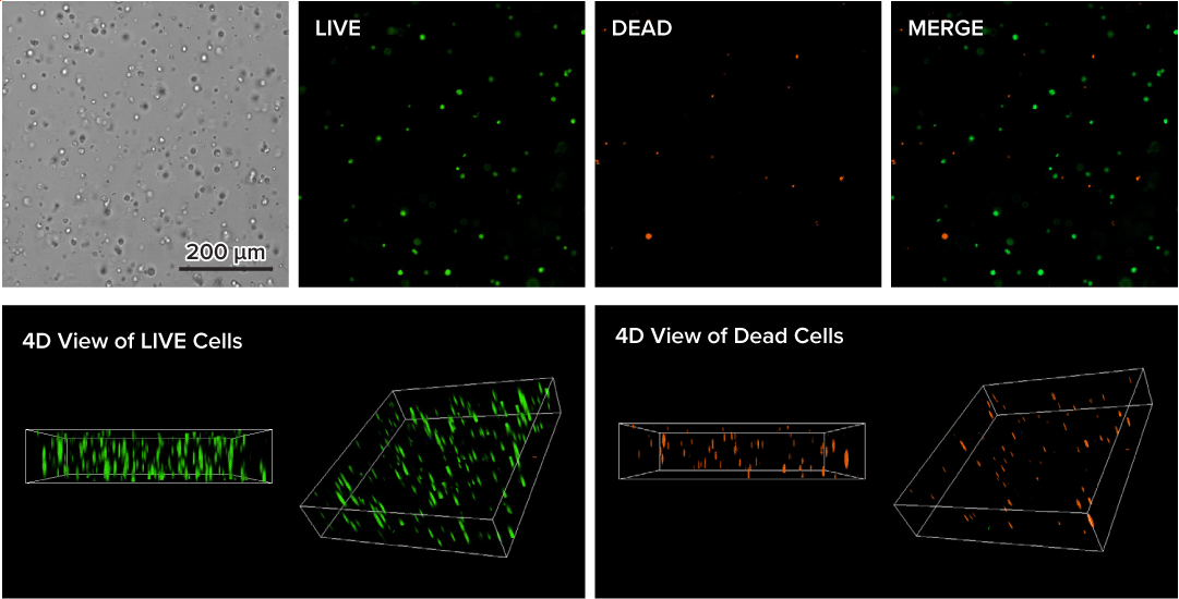
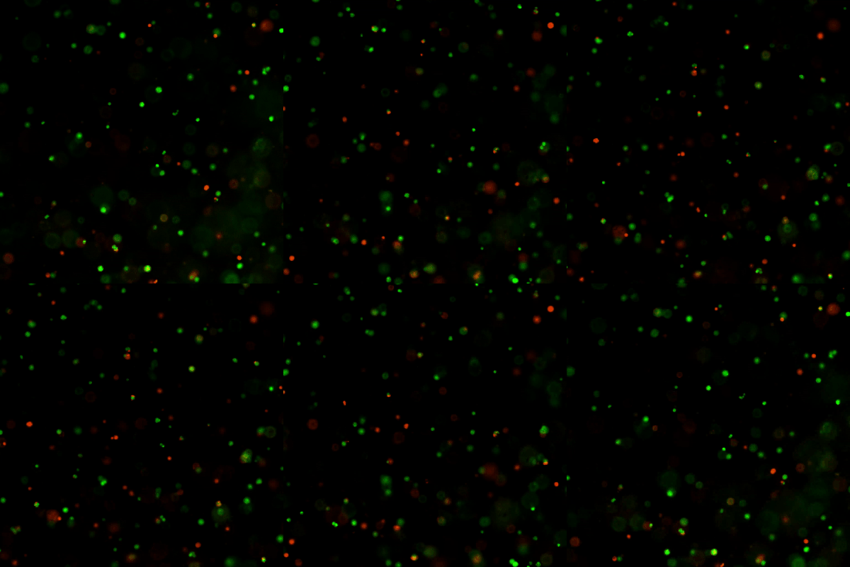
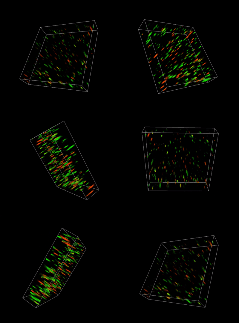
Figure 3. Live-dead cell viability analysis by using Cyto3D® Live-Dead Assay Kit.
Glioblastoma cells (SF 298, about 60% cell viability) were 3D cultured in VitroGel® system for 2 days. Two microliters (2 µL) of Cyto3D® Live-Dead Assay reagent was added to each well containing 50 µL hydrogel and 50 µL cover medium. The mixture was incubated at 37°C for 5-10 min. The cells were then observed under a fluorescence microscope. The images show the Live (green) and Dead (orange) cells within the 3D hydrogel matrix. The z-stack images of cells within hydrogel were then 3D reconstructed and shown in the 4D view images. The live and dead cells at higher levels of the hydrogel are clearly shown in the images using the Cyto3D® Live-Dead Assay Kit.
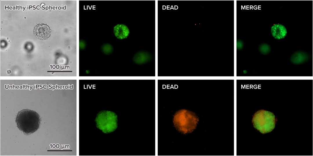
Figure 4. Live-dead cell viability images of stem cell spheroids.
Stem cells were static suspension-cultured in VitroGel® STEM (CAT# VHM02) for 5 days. Two microliters (2 µL) of Cyto3D® Live-Dead Assay reagent was added to each well containing 100 µL cell suspension. The mixture was incubated at 37°C for 5-10 min. The cells were then observed under a fluorescence microscope. The images show the Live (green) and Dead (orange) stem cell spheroids cultured in a 3D hydrogel matrix. The live-dead dyes of Cyto3D® Live-Dead Assay Kit can successfully penetrate into large cell spheroids for cell viability analysis.Data and References

Figure 1: Live-dead cell viability images: Intestinal organoids stained with Cyto3D® Live-Dead Assay Kit.
Intestinal organoids were cultured in regulated conditions for 5 days. Six microliters of Cyto3D® reagent were mixed with organoid culture media (each well includes 150 µL of organoid culture media and 150 µL of hydrogel volume). The mixture was incubated at 37°C for 10-15 min, and the cells were observed under a fluorescence microscope. (A) A bright field image of a mature intestinal organoid. Images show live cells (B: Green) and dead cells (C: Red) in a mature intestinal organoid.

Figure 2: Live-dead cell viability images: Intestinal organoids stained with Cyto3D® Live-Dead Assay Kit.
Intestinal organoids were cultured in regulated conditions for 2-3 days. Six microliters of Cyto3D® Live-Dead Assay reagent were mixed with organoid culture media (each well includes 150 µL of organoid culture media and 150 µL of hydrogel volume). The mixture was incubated at 37°C for 10-15 min, and the cells were observed under a fluorescence microscope. (A) A brightfield image of a young healthy intestinal organoid. Images show live cells (B: Green) and dead cells (C: Red) in a healthy intestinal organoid.


Figure 3. Live-dead cell viability analysis by using Cyto3D® Live-Dead Assay Kit.
Glioblastoma cells (SF 298, about 60% cell viability) were 3D cultured in VitroGel® system for 2 days. Two microliters (2 µL) of Cyto3D® Live-Dead Assay reagent was added to each well containing 50 µL hydrogel and 50 µL cover medium. The mixture was incubated at 37°C for 5-10 min. The cells were then observed under a fluorescence microscope. The images show the Live (green) and Dead (orange) cells within the 3D hydrogel matrix. The z-stack images of cells within hydrogel were then 3D reconstructed and shown in the 4D view images. The live and dead cells at higher levels of the hydrogel are clearly shown in the images using the Cyto3D® Live-Dead Assay Kit.

Figure 4. Live-dead cell viability images of stem cell spheroids.
Stem cells were static suspension-cultured in VitroGel® STEM (CAT# VHM02) for 5 days. Two microliters (2 µL) of Cyto3D® Live-Dead Assay reagent was added to each well containing 100 µL cell suspension. The mixture was incubated at 37°C for 5-10 min. The cells were then observed under a fluorescence microscope. The images show the Live (green) and Dead (orange) stem cell spheroids cultured in a 3D hydrogel matrix. The live-dead dyes of Cyto3D® Live-Dead Assay Kit can successfully penetrate into large cell spheroids for cell viability analysis.
